[ Ana Sayfa | Editörler | Danışma Kurulu | Dergi Hakkında | İçindekiler | Arşiv | Yayın Arama | Yazarlara Bilgi | E-Posta ]
Fırat Tıp Dergisi
2008, Cilt 13, Sayı 1, Sayfa(lar) 043-048
[ Özet ] [ PDF ] [ Benzer Makaleler ] [ Yazara E-Posta ] [ Editöre E-Posta ]
Effect of Inhaled N-Acetyl-L-Cysteine Treatment on Induced Sputum Glutathione and Nitrite
Figen DEVECİ1, Teyfik TURGUT1, Nevin İLHAN2, Gamze KIRKIL1, Mehmet Hamdi MUZ1
1Fırat Üniversitesi, Tıp Fakültesi Göğüs Hastalıkları Anabilim Dalı, ELAZIĞ
2Fırat Üniversitesi, Tıp Fakültesi Tıbbi Biyokimya Anabilim Dalı, ELAZIĞ
Keywords: Induced sputum, glutathione, nitrit, exacerbation of asthma, N-acetyl-L-cysteine, İndükte balgam, glutatyon, nitrit, astım atağı, N-asetil-L-sistein
Summary
Objective: An imbalance between oxidants-antioxidants is known to play an important role in the pathogenesis of asthma, especially during exacerbation. The aim of this study was to investigate the effect of N-acetyl-L-cysteine (NAC) treatment on the levels of sputum nitrit (NO2-) and reduced glutathione (GSH) contents in patients with asthma during exacerbations.

Materials and Methods: The study had a double blind, placebo controlled design. Induced sputum GSH, NO2 -, cell counts and plasma NO2 contents were evaluated in 11 healthy controls (HCs) and 25 patients with asthma during exacerbation before and after treatment. Fifteen patients with acute asthma attack were treated with inhaled NAC during exacerbation.

Results: Plasma-sputum NO2 - and sputum GSH contents were significantly higher in subjects with asthma subgroups than HCs (p<0.01 for NAC treatment group, p<0.005 for placebo treatment group, for plasma NO2 -; p<0.001 for both groups for sputum NO2 -; p<0.001 for both groups for sputum GSH). NAC treatment group had higher GSH contents in sputum samples after treatment which were, however, not significantly different from those before treatment. There was a significant decrease in sputum (p<0.005 for NAC treatment group, p<0.01 for placebo group) and plasma NO2 - (p<0.005 for NAC treatment group, p<0.01 for placebo group) levels in both NAC treatment group and placebo group after treatment when compared with the pretreatment levels.

Conclusion: These findings indicated that addition of NAC to standard attack therapy does not modify the oxidant-antioxidant status in asthma patients during exacerbation.©2008, Firat University, Medical Faculty.

  • Top
  • Summary
  • Introduction
  • Methods
  • Results
  • Disscussion
  • References
  • Introduction
    It has been recognized for many years that patients who die from acute asthma attacks have grossly inflammed airways. As inflammation is often associated with an increased generation of reactive oxygen species (ROS), and the biochemical environment in the asthmatic airways is favourable for free radical mediated reactions, it is rational to surmise that an oxidant stress could be mechanistically important in asthma1. An imbalance between oxidants and antioxidants, in favor of oxidants leading to oxidative stress, is known to play an important role in the pathogenesis of asthma, and increased oxidative stress is related to disease severity and may amplify the inflammatory response, particularly during exacerbations2,3. Most studies using blood leukocytes and cells isolated from bronchoalveolar lavage (BAL) of asthmatic patients indicate enhanced ROS generation compared with control subjects4,5 and decreased antioxidant capacity in plasma of asthmatic patients, both in stable and in acute asthma6. We have previously demonstrated increased oxidant capacity in sputum of both stable and exacerbations of asthma and decreased antioxidant capacity in exacerbations of asthma compared with stable phase7.

    Despite the abundance of evidence indicating elevated oxidative stress and reduced antioxidant defences in asthma, antioxidant supplementation studies to date have been controversial. N/acetyl-L-cysteine (NAC) is a thiol containing compound that is used to reduce viscosity and elasticity of mucus. It is able to scavenge hydrogen peroxide, hydroxyl radicals and hypochlorous acid8. NAC can be deacetylated to cysteine, an important procursor of cellular GSH sythesis, and thus stimulating the cellular GSH system9. In view of this, we wondered whether inhaled NAC might also be useful in increasing anti-oxidant capacity during exacerbation. We undertook a double blind, placebo controlled study of inhaled NAC 600 mg bid in addition to standard treatment in patients admitted to hospital with an acute execerbation of asthma.

  • Top
  • Summary
  • Introduction
  • Methods
  • Results
  • Disscussion
  • References
  • Methods
    Study Design
    The study had a double blind, placebo controlled design. The study was performed in asthma patients who were admitted and hospitalized to our clinic for asthma exacerbation between January-April 2005. Patients were immediately evaluated and studied before any asthma treatment. Subjects had mild to moderate exacerbation of acute asthma. Acute asthma patients who had severe exacerbation were excluded from this study. Patients had no systemic diseases, malignancy, vascular disease, thrombosis, alcoholism, renal disease and hepatic disease. Their therapy was done according to the international asthma guidelines10. Patients who received oral/parenteral corticosteroids, antibiotics, theophylline and antioxidant vitamins were excluded from the study. Hospitalized patients are taken randomly and were separated into two groups. NAC treatment group received nebulized NAC (15 acute asthma patients, twice a day, 300 mg ampuls) and placebo treatment group received nebulized 0.9% NaCl ampul (10 acute asthma patients, twice a day) during exacerbation. The patients were reevaluated 15 days after the therapy.

    Patients/Subjects
    Acute asthma patients Twenty-five non-smoking atopic asthma patients during exacerbation participated in the study. All subjects were atopic with positive skin prick testing for common aeroallergens from our area (house-dust mites, grass pollen, cat and dog dander, mould mixture). Positive skin prick test was defined when wheal was 3 mm when compared to salin. The diagnosis of asthma was based on international guidelines10. An exacerbation of asthma was defined by the presence of the dyspnea at rest with wheezing or nocturnal symptoms disturbing sleep. Their current therapy (before the exacerbation) was shown in Table 1.
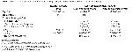

    Click Here to Zoom
    Table I: Demographics and lung function datas of all groups, and current medications of asthma subgroups

    “Healthy controls” (HCs) Eleven age-matched, nonsmoking healthy subjects were included as control subjects. All subjects were randomly selected from hospital staff. Inclusion criteria for non-smokers were; no history of respiratory or allergic disease, normal baseline spirometric parameters as predicted for age, sex and height, no history of upper respiratory tract infection in the preceeding 6 weeks, and no use of any regular medication. The Ethics Committe of Firat University Faculty of Medicine reviewed and approved the protocol, and all subjects gave informed consent to participate in the study.

    Pulmonary Function Test
    Pulmonary function parameters (FEV1, FVC, FEF25-75) were measured with a spirometer (SuperSpiro, Micromedical Limited, England).

    Sputum Induction
    Sputum was induced during the acute exacerbation as previously described11. Sputum induction was performed by inhalation of 3% NaCl for 20 minutes from nebuliser [Porta- Neb compressor, Medic-Aid Sidestream nebuliser chamber, mass median diameter 3.18 m (Medic-Aid Limited, UK)]. The standard safety precaution was to premedicate with 200 Ug of inhaled salbutamol12. Before and after sputum induction lung function measurements were performed. The safety of sputum induction was monitored by measuring peak expiratory flow rates (PEFR). The procedure would have been stopped if PEFR were decreased by 25%. The sputum induction procedure did not cause troublesome symptoms and the PEFR was not decreased by more than 25% in all “acute asthma patients”. Treatment of exacerbation was made according to international guidelines in all acute asthma patients after sputum induction. Expectorated sputum was collected in sterile plastic tubes placed on ice to slow down metabolic processes that might result in loss of GSH.

    Sample Processing
    Sputum samples were processed within 30 minutes of collection using the method described by Dauletbaev et al11. Samples were diluted with three volumes of chilled phosphate buffered saline (PBS: all reagents were purchased from Sigma- Aldrich Chemie GmbH, Steinheim, Germany). Supernatants were obtained by centrifugation (300 g, 15 minutes, 4ºC) and transferred to another vial by filtering through multiple layers of cotton gauze. Additional centrifugation (800 g, 5 minutes, 4ºC) ensured removal of the remaining cell debris and mucus. Aliquots of the supernatants were placed on ice and assayed immediately for reduced glutathione supernatant was waited at -20 ºC for measuring nitrit (NO2 -) contents.

    GSH Measurement
    The sputum GSH was measured using an enzymatic recycling assay13,14. The standard and sample solutions were added to an equal volume of DTNB and 50 µl of this mixture (final concentration of DTNB 0.25 mM) were pipetted into a 1 ml cuvette followed by glutathione reductase and NADPH (final concentrations 1 U/ml and 0.22 µM, respectively). The reaction mixture was equilibrated and the kinetic reaction was followed for two minutes at 412 nm (Techcomp Ltd., UV-VIS 8500 spectrophotometer, Hong Kong).

    NO2 Measurement
    The measurement of plasma and supernatant NO was difficult because this radical was poorly soluble in water and had a short half-life in tissue (10-60 s), but its half-life might be as long as 4 min in the presence of oxygen. For these reasons, the determination of NO itself was difficult and required the handling of radioisotopes. In spite of this, the end products, nitrate and nitrite, were preferentially used in clinical biochemistry. Nitrite, a stable end -product of NO, was measured in plasma by using the spectrophotometric Griess reaction15. One thousand mL experimental samples of deproteinised plasma was reacted with 500 mL N naphthylethylenediamine, 10 g/L sulfanilamide for 45 min at room temperature and analyzed by spectrophotometry at 545 nm. Concentrations were determined by comparison with sodium nitrite. The lower limit of detection was 0.2 mmol/L.

    Statistical Analysis
    All statistical analyses were done using SPSS v10.0 software. Data were expressed as mean ± standard deviation (SD). Statistical analysis was performed using Kruskal-Wallis test for multiple-group comparisons; Mann-Whitney U test was performed to test any observed differences for significance. Wilcoxon’s rank sum test was performed for comparisons before and after treatment values of asthma subgroups. Chisquare test was performed to compare gender distrubion between groups. A p value of <0.05 was considered as statistically significant.

  • Top
  • Summary
  • Introduction
  • Methods
  • Results
  • Disscussion
  • References
  • Results
    There was no significant difference in mean age and sex between healthy subjects, and both asthma subgroups. The mean FEV1 levels were significantly lower in both asthma subgroups compared with healthy controls (p<0.001 for both group), but there was no significant difference between asthma subgroups. There was no significant difference in mean duration of disease between asthma subgroups. A small decrease in lung function parameters was observed during the sputum induction procedure and there was no significant difference in FEV1 between the groups. Patients’ characteristics, current medications are given in Table I.

    At exacerbation, sputum GSH contents were higher in both acute asthma subgroups before treatment than HCs (p<0.001 for both groups), and remained higher also after treatment in both acute asthma groups (p<0.001 for both groups). There was no significant difference betweenacute asthma subgroups in sputum GSH levels before and after treatment. There was no significant difference in sputum GSH levels in both acute asthma subgroups after treatment when compared with the pretreatment levels, although there was a trend toward higher levels in patients who received NAC (Table II).


    Click Here to Zoom
    Table II: Comparison of sputum cell counts, GSH, NO2 - and plasma NO2 - levels from asthma patients during exacerbations and healthy control subjects

    Sputum NO2 - contents were higher in both acute asthma subgroups before treatment than HCs (p<0.001 for both groups). After treatment, sputum NO2 - contents were significantly lower in placebo group when compared to HCs (p<0.05) but there was no significant difference between NAC treatment group and HCs. There was no significant difference between acute asthma subgroups in sputum NO2 - contents before and after treatment. We observed a significant decrease in sputum NO2 - levels in both acute asthma subgroups after treatment (p<0.005 for NAC treatment group, p<0.01 for placebo treatment group) when compared with the pretreatment levels (Table II).

    Before treatment, plasma NO2 - contents were higher in both acute asthma subgroups than HCs (p<0.01 for NAC treatment group, p<0.005 for placebo treatment group), but there was no significant difference between acute asthma groups. After therapy, there was no significant difference in plasma NO2 - contents between all groups. There was a significant difference in plasma NO2 - contents in both acute asthma subgroups after treatment when compared with the pretreatment levels (p<0.005 for NAC treatment group, p<0.01 for placebo treatment group) (Table II).

    FEV1 was significantly higher in both asthma subgroups after treatment than those before treatment (p<0.005 for NAC treatment group, p<0.01 for placebo treatment group). There was no significant difference in FEV1 between NAC treatment group and placebo treatment group after treatment and the rate of change in FEV1 was greater with NAC than with placebo but this difference was not statistically significant (Table II).

    Sputum GSH, NO2 - and plasma NO2 - contents of both asthma subgroups before and after treatment were shown in figure 1 and 2.


    Click Here to Zoom
    Figure 1: Sputum GSH, NO2 -, plasma NO2 - contents in patients who received NAC treatment


    Click Here to Zoom
    Figure 2: Sputum GSH, NO2 -, plasma NO2 - contents in patients who received placebo treatment

  • Top
  • Summary
  • Introduction
  • Methods
  • Results
  • Disscussion
  • References
  • Discussion
    This study showed that sputum GSH and NO2 - contents were increased in asthma during exacerbation when compared with HCs and sputum GSH levels were slightly modified during acute asthma attack therapy with inhaled NAC. In addition, sputum and plasma NO2 - contents were decreased after treatment in all asthma patients.

    GSH is the key antioxidant in the respiratory tract lining fluid (RTLF). Disturbed GSH status is reported in asthma patients. Kelly and Smith obtained an increased GSH (total and oxidised) status in BAL fluid in asthma patients 16,17. Doulatbaev et al also demonstrated that stable asthma patients tended to have higher GSH concentrations in sputum samples11. There are several clinical studies of the oxidantantioxidant status in asthma. Acute exacerbations of these conditions have received much less attention. We hypothesised that increased numbers of inflammatory leukocytes in the bronchoalveolar space and/or those sequestered in the pulmonary vasculature in acute exacerbations of asthma, may create an increased oxidant burden. Rahman et al showed that decreased antioxidant and increased oxidant capacity in plasma of patients presenting with acute exacerbations of asthma, and antioxidant capacity tended to improve by the time of discharge6. We observed increased amount of sputum GSH in patients with asthma during exacerbation. This result suggests that GSH synthesis and/or transport were increased in response to the presence of excess oxidants during acute exacerbation. A compensatory but probably inadequate increase in GSH has been reported in other conditions of oxidative stress18,19. If ROS and RNS are important in asthma especially during acute exacerbation, enhancement of the antioxidant defences would be expected to have beneficial effects in the disaese. On the contrary, some studies evaluated GSH of exhaled breath condensate in children with asthma during acute exacerbations were found decreased GSH levels compared with control subjects and stable asthma patients20,21. At present, it is not clear whether altered RTLF GSH status is the cause or a consequence in respiratory disease. Studies carried out with normal volunteers showed that GSH and other antioxidants present in RTLF play an important role in protecting the lung surface from oxidative attack22.

    Sputum concentrations of NO2 - and nitrate, which are stable metabolites of NO have been shown to be elevated in asthma23,24. NO can be measured in the exhaled air of asthmatic patients25,26, and it is thought to reflect the inflammatory state of the airways. In the current study, we demonstrated that sputum NO2 - content was higher in acute asthma exacerbation when compared with the healthy subjects and their levels were significantly decreased after treatment. To our knowledge, this is the first study in which sputum NO2 - contents in acute asthma exacerbation were evaluated, therefore the data cannot be compared with previous findings. Recently, Hunt et al showed that the pH in the airways drops dramatically during acute asthma attack, which facilitates the conversion of nitrite to NO. Hence, increased NO concentrations in the exhaled air of asthmatic patients may reflect nitrite conversion rather than NOS activity. Acidosis at levels seen in subjects with acute asthma causes both necrosis of human eosinophils and conversion of airway NO2 - to NO effects relevant to asthma pathophysiology27. In addition, increased sputum NO2 - contents may develop due to increased numbers of inflammatory leukocytes in the bronchoalveolar space during acute exacerbations of asthma. High concentrations of NO released in an inflammatory context, it has putatively detrimental effects via the formation of ROS28.

    Despite the abundance of evidence indicating elevated oxidative stress and reduced antioxidant defences in asthma, antioxidant supplementation studies to date have been limited. NAC is one of the most widely investigated antioxidants having shown beneficial effects in disease states. In contrast with the activity found different pulmonary disease, the influence of NAC in clinical asthma remains uncertain29,30 but has not been recently evaluated. We found that short-term administration of inhaled 600 mg/day NAC increased sputum GSH contents in acute asthma patients, but this increase was not significant according to before treatment. When compared with placebo group, NAC group had increased sputum GSH levels after treatment but it was not significant. Administration of NAC had no significant effect on FEV1, serum and plasma NO2 -, and sputum GSH levels in asthma patients during exacerbations. Improvement in postreatment levels of FEV1 could be attributed to the use of standard attack therapy (Â2 agonists and/or inhaled steroids). NAC can easily be deacetylated to cysteine, an important precursor of cellular GSH synthesis, and thus stimulate the cellular GSH system9. The upper level of cellular GSH is regulated by feedback inhibition, but cellular levels also depend upon availability of the substrates, especially cysteine31. In several studies, it was shown that intracellular and plasma cysteine levels were increased following NAC administration32,33. Meyer et al found that GSH levels increased in BAL fluid after the oral NAC (3x600 mg NAC per day for 5 days) augmentation compared with pretherapy34. On the contrary, Cotgreave et al demonstrated that the cysteine and GSH content in the lavage fluid were unaltered during NAC (600 mg, daily for 2 weeks) administration in healthy subjects35. NAC in a daily dose up to 1800 mg administered for 5 days did not significantly increase the concentration of GSH or cysteine in BAL fluid36. Oral administration of NAC for several days did not elevate the concentration of GSH in the airways. There has been many studies about oral administration of NAC but a few studies is present with nebulized NAC administration. In these studies it is shown that inhaled NAC has anti- or prooxidant action37 and it is reported that nebulized NAC transiently increases exhaled H2O2 level, whereas it has no effect on other oxidative parameters38. In our study, sputum GSH contents were not increased significantly in patients with acute asthma attacks. This may be due to both short-term and nebulized administration of NAC during exacerbation.

    In conclusion, the results of this study demonstrate that short-term inhaled treatment with NAC produced insufficient increase in sputum GSH contents in the airways in patients with asthma during exacerbation. In addition, this therapy had no effect on sputum NO2 - content. For this reason, our study does not suggest that NAC supplementation therapy is effective during asthma exacerbations. Further studies can be made by using higher doses of NAC to mild to moderate asthma patients.

  • Top
  • Summary
  • Introduction
  • Methods
  • Results
  • Discussion
  • References
  • References

    1) Dworski R. Oxidant stress in asthma. Thorax 2000; 55: 51-53.

    2) Barnes PJ. Pathophysiology of asthma. In: Asthma. Chung F, Fabbri LM (eds). The European Respiratory Monograph, ERS Journals Ltd, Monograph 2003; 23: 84-113.

    3) Owen S, Pearson D, Suarez-Mendez V, et al. Evidence of freeradical activity in asthma. N Engl J Med 1991; 325: 586-587.

    4) Calhoun WJ, Bush RK. Enhanced reactive oxygen species metabolism of airspace cells and airway inflammation follow antigen challenge in human asthma. J Allergy Clin Immunol 1990; 86: 306-313.

    5) Henrichs PAJ, Nijkamp FP. Reactive oxygen species as mediators in asthma. Pulmonary Pharmacology and Therapeutics 2001; 14: 409-421.

    6) Rahman I, Morrison D, Donaldson K, Mac Nee W. Systemic oxidative stress in asthma, COPD, and smokers. Am J Respir Crit Care Med 1996; 154: 1055-1060.

    7) Deveci F, Ilhan N, Turgut T, et al. Glutathione and nitrite in induced sputum from patients with stable and acute asthma compared with controls. Ann Allergy Asthma Immunol 2004; 93: 91-97.

    8) Aruoma OI, Halliwell B, Hoey BM, Butler J. The antioxidant action of N-acetylcysteine: its reaction with hydrogen peroxide, hydroxyl radical, superoxide, and hypochlorous acid. Free Radic Biol Med 1989; 6: 593-597.

    9) Gillissen A, Nowak D. Characterization of N-acetylcysteine and ambroxol in antioxidant therapy. Respir Med 1998; 92: 609-623.

    10) National Heart, Lung, and Blood Institute, National Institutes of Health. Global Strategy for Asthma Management and Prevention NHLBI/WHO Workshop Report Revised Global Initiative for Asthma (GINA 2002), Publication no. 02-3659. Bethesda: National Institutes of Health, 2002.

    11) Dauletbaev N, Rickmann J, Viel K, et al. Glutathione in induced sputum of healthy individuals and patients with asthma. Thorax 2001; 56: 13-18.

    12) Vlachos-Mayer H, Leigh R, Sharon RF, et al. Success and safety of sputum induction in the clinical setting. Eur Respir J 2000; 16: 997-1000.

    13) Akerboom TP, Sies H. Assay of glutathione, glutathione disulfide, and glutathione mixed disulfides in biological samples. Methods Enzymol 1981; 77: 373-382.

    14) Buhl R, Vogelmeier C, Critenden M, et al. Augmentation of glutathione in the fluid lining the epithelium of the lower respiratory tract by directly administering glutathione aerosol. Proc Natl Acad Sci U S A 1990; 87: 4063-4067.

    15) Green LC, Wagner DA, Glogowski J, et al. Analysis of nitrate, nitrite and [15N] nitrate in biological fluids. Anal Biochem 1982; 126: 131-138.

    16) Kelly FJ, Mudway I, Blomberg A, et al. Altered lung antioxidant status in patients with asthma. Lancet 1999; 354: 482-483.

    17) Smith LJ, Houston M, Anderson J. Increased levels of glutathione in bronchoalveolar lavage from patients with asthma. Am Rev Respir Dis 1993; 147: 1461-1464.

    18) Borm PJ, Bast A, Wouters EF, et al. Red blood cell antioxidant parameters in healthy elderly control subjects versus silicosis patients. Free Rad Res Comm 1987; 3: 117-127.

    19) Toth KM, Berger EM, Beehler CJ, Repine JE. Erytrocytes from cigarette smokers contain more glutathione and catalase and protect endothelial cells from hydrogen peroxide than do erytrocytes from nonsmokers. Am Rev Respir Dis 1986; 134: 281- 284.

    20) Bibi H, Schlesinger M, Tabachnic E, et al. Erytrocyte glutathione peroxidase activity in asthmatic children. Ann Allergy 1988; 61: 339-340.

    21) Corradi M, Folesani G, Andreoli R, et al. Aldehydes and glutathione in exhaled breath condensate of children with asthma exacerbation. Am J Respir Crit Care Med 2003; 167: 395-399.

    22) Kelly FJ. Glutathione: in defence of the lung. Food and Chemical Toxicology 1999; 37: 963-966.

    23) Gustafsson L. Exhaled nitric oxide as a marker in asthma. Eur Respir J 1998; 11 (Suppl. 26): 49-52.

    24) Kanazawa H, Shoji S, Yamada M, et al. Increased levels of nitric oxide derivatives in induced sputum in patients with asthma. J Allergy Clin Immunol 1997; 99: 624-629.

    25) Dupont LJ, Rochette F, Demedts MG, Verleden GM. Exhaled NO correlates with airway hyperresponsiveness in steroid-naive patients with mild asthma. Am J Respir Crit Care Med 1998; 157: 894-898.

    26) Massaro AF, Mehta S, Lilly CM, et al. Elevated NO concentrations in isolated lower airways gas of asthmatic subjects. Am J Respir Crit Care Med 1996; 153: 1510-1514.

    27) Hunt JF, Fang K, Malik R, et al. Endogenous airway acidification. Implications for asthma pathophysiology. Am J Respir Crit Care Med 2000; 161: 694-699.

    28) Beckman JS, Koppenol WH. NO, superoxide, and peroxynitrite: the good, the bad, and the ugly. Am J Physiol 1996; 40: 1424- 1437.

    29) Bylin G, Hedenstierna G, Lagerstrand L, Wagner PD. No influence of acetylcysteine on gas exchange and spirometry in chronic asthma. Eur J Respir Dis 1987; 71: 102-107.

    30) Millman M, Millman FM, Goldstein IM, Mercandetti AJ. Use of acetylcysteine in bronchial asthma--another look. Ann Allergy 1985; 54: 294-296.

    31) Meister A. Glutathione metabolism and its selective modification. J Biol Chem 1988; 263: 17205-17208.

    32) Bridgeman MM, Marsden M, MacNee W, et al. Cysteine and glutathione concentrations in plasma and bronchoalveolar lavage fluid after treatment with N-acetylcysteine. Thorax 1991; 46: 39- 42.

    33) Mulier B, Rahman I, Watchorn T, et al. Hydrogen peroxideinduced epithelial injury: the protective role of intracellular nonprotein thiols (NPSH). Eur Respir J 1998; 11: 384-391.

    34) Meyer A, Buhl R, Magnussen H. The effect of oral Nacetylcysteine on lung glutathione levels in idiopathic pulmonary fibrosis. Eur Respir J 1994; 7: 431-436.

    35) Cotgreave IA, Eklund A, Larsson K, Moldeus PW. No penetration of orally administered N-acetylcysteine into bronchoalveolar lavage fluid. Eur J Respir Dis 1987; 70: 73-77.

    36) Bridgeman MM, Marsden M, Selby C, et al. Effect of N-acetyl cysteine on the concentrations of thiols in plasma, bronchoalveolar lavage fluid, and lung tissue. Thorax 1994; 49: 670-675.

    37) Szkudlarek U, Zdziechowski A, Witkowski K, et al. Effect of inhaled N-acetylcysteine on hydrogen peroxide exhalation in healthy subjects. Pulm Pharmacol Ther 2004;17: 155-162.

    38) Rysz J, Stolarek RA, Luczynski R, et al. Increased hydrogen peroxide concentration in the exhaled breath condensate of stable COPD patients after nebulized N-acetylcysteine. Pulm Pharmacol Ther 2007; 20: 281-289.

  • Top
  • Summary
  • Introduction
  • Methods
  • Results
  • Discussion
  • References
  • [ Başa Dön ] [ Özet ] [ PDF ] [ Benzer Makaleler ] [ Yazara E-Posta ] [ Editöre E-Posta ]
    [ Ana Sayfa | Editörler | Danışma Kurulu | Dergi Hakkında | İçindekiler | Arşiv | Yayın Arama | Yazarlara Bilgi | E-Posta ]