[ Ana Sayfa | Editörler | Danışma Kurulu | Dergi Hakkında | İçindekiler | Arşiv | Yayın Arama | Yazarlara Bilgi | E-Posta ]
Fırat Tıp Dergisi
2008, Cilt 13, Sayı 1, Sayfa(lar) 049-052
[ Özet ] [ PDF ] [ Benzer Makaleler ] [ Yazara E-Posta ] [ Editöre E-Posta ]
Determination of the Role of Mean Corpuscular Volume Level on the Diagnosis of Alcoholic Liver Cirrhosis and Investigation of Its Effect on the Prognosis
Özgür TANRIVERDİ1, Esen GER2, Sernaz UZUNOĞLU2, M. Kemal SEREZ2, B. Ümit ÜRE2, Kadir ERGEN2, A.Baki KUMBASAR2
1Goztepe Safak Private Hospital, Internal Medicine and Emergency Service, İSTANBUL
2Haseki Education and Research Hospital, 3rd Internal Medicine Clinic, İSTANBUL
Keywords: Cirrhosis, alcohol, macrocytosis, prognosis, Siroz, alkol, makrositoz, prognoz
Summary
Objectives: In this study, we aimed to determine the relationship between macrocytosis and Alcoholic Liver Cirrhosis (ALC) and its accuracy, sensitivity and specificity rates.

Material and Methods: Mean corpuscular volume (MCV) levels of 46 patients diagnosed as having ALC (Group 1) and 51 patients diagnosed as having Hepatitis B cirrhosis and Hepatitis C cirrhosis (Group 2) were compared retrospectively.

Results: MCV level determined as 94.6±10.87 in patients in Group 1 was statistically significantly higher than in patients in Group 2 (p<0.001). Macrocytosis was determined in 26 patients (56.52%) in Group 1 but in 3 patients (5.88%) in Group 2. When the cut-off value for MCV in the diagnosis of ALC was taken as 102 fl, its accuracy, sensitivity and specificity rates for the diagnosis were determined as 78%, 93% and 72% respectively. MCV levels of Child class C ALC patients (n=27) were determined statistically significantly higher than MCV levels of Child class A and Child class B ALC patients.

Conclusion: We concluded that MCV level is an important variable for the diagnosis of ALC but it is insufficient alone for the diagnosis; macrocytosis frequency increases in these patients and MCV levels increase as the prognosis worsens even without existence of anemia. ©2008, Firat University, Medical Faculty

  • Top
  • Summary
  • Introduction
  • Methods
  • Results
  • Disscussion
  • Conclusion
  • References
  • Introduction
    Whether anemia exists or not, some changes in erythrocyte morphology are observed in patients diagnosed as Chronic Liver Disease (CLD). These changes can be related with erythrocyte membrane morphology or erythrocyte volume1. When mean corpuscular volume (MCV) and erythrocyte morphology are evaluated, it is shown in many studies that normocytic and macrocytic morphology are observed in especially anemic patients diagnosed as having chronic liver disease (CLD)1. It is determined in many other studies that macrocytic morphologic changes are seen especially in patients diagnosed as having alcoholic liver disease (ALD) with or without anemia2-7.

    We aimed in our study to investigate and show whether the MCV in patients diagnosed as having alcoholic liver cirrhosis(ALC) is in harmony with macrocytosis degree or not and whether it is so different from MCV level in patients diagnosed as having non-alcoholic liver cirrhosis or not.

  • Top
  • Summary
  • Introduction
  • Methods
  • Results
  • Disscussion
  • Conclusion
  • References
  • Methods
    Patients who attended to 3rd Internal Medicine policlinic, 3rd Internal Medicine clinic and hepatology policlinic of Haseki Education and Research Hospital between October 1998 and July 2002 were examined retrospectively and included to our study. Totally 97 patients with liver cirrhosis included to our study were divided in to two groups according to etiologies. 46 patients who has diagnosed as having ALC were classified as Group 1 and 51 patients diagnosed as having Hepatitis B cirrhosis and Hepatitis C cirrhosis were classified as Group 2. Anemia wasn’t included to our study as a criterion. Whole blood counts obtained from all patients by automatic analyzers were examined and MCV levels were recorded. The relationship between the existence of ALC and MCV was evaluated. The cut-off value for MCV was determined and its accuracy, sensitivity and specificity rates for the diagnosis of ALC were calculated and examined. Accuracy, sensitivity and specificity values were calculated according to the formulas shown in Table 1.


    Click Here to Zoom
    Table 1: The formulas of truth, sensitivity and specificity a/a+c for sensitivity, d/b+d for specificity and a+d/N for accuracy

    The relationship between the MCV and the prognosis of liver cirrhosis (LC) that is another aim emphasized in our study is evaluated by considering the Child-Pugh criteria (a classification used to classify the prognosis of LC). Child-Pugh criteria determining the prognosis in patients with LC are shown in Table 2.


    Click Here to Zoom
    Table 2: Child-Pugh classification and point degree Child-Pugh grade A; 5-6 point Child-Pugh grade B 7-9 point Child-Pugh grade C 10-15 point

    The statistical evaluation was performed by using for SPSS 9.0 programme for the Windows. Student-t test, chisquare test and Pearson correlation test were used for statistical significance.

  • Top
  • Summary
  • Introduction
  • Methods
  • Results
  • Disscussion
  • Conclusion
  • References
  • Results
    The demographic features of patients included in our study are shown in Table 
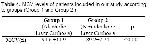
3, MCV levels of patients included in our study according to groups (Group 1 and Group 2) are shown in Table 4 and the distributions of Formulas shown in Table 
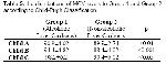
1 were used to calculate accuracy, sensitivity and specificity rates. MCV levels to Group 1 and Group 2 according to Child-Pugh Classification are shown in Table 5.


    Click Here to Zoom
    Table 3: the demographic features of patients included in our study


    Click Here to Zoom
    Table 4: MCV levels of patients included in our study according to groups (Group 1 and Group 2 )


    Click Here to Zoom
    Table 5: the distributions of MCV levels to Group 1 and Group 2 according to Child-Pugh Classification

    The study variables of patients included in our study were investigated independently from gender and age features. In our study, the MCV level of patients in Group 1 were found 94.6 ±11.9 and this was statistically significantly higher than MCV level of patients in Group 2 (p<0.001).

    When the relationship between the Child-Pugh Classification and the MCV level was investigated in patients diagnosed as having ALC (Group 1), it was determined that MCV level was statistically significantly higher in Child class C patients than Child class A and B patients (respectively; p<0.001, p<0.01). Furthermore, the MCV levels of Child B and Child C class alcoholic liver cirrhosis patients were statistically significantly higher than MCV levels of patients in all Child classes (Child A, B, C) in Group 2 (p<0.001, p<0.01, p<0.01 ve4 p<0.05, p<0.01, p<0.05 respectively). Similarly, when the Child A, B, C class patients in Group 1 and Group 2 were compared among themselves no statistically significant difference was determined between Child A class patients in Group 1 and Class A patients in Group 2 in respect of MCV level (p>0.01). A statistically significant difference was determined between Child class B patients in Group 1 and Child class B patients in Group 2 and between Child class C patients in Group1 and Child class C patients in Group 2 in respect of MCV level (p<0.001, p<0.05 respectively)

    Macrocytosis frequency in patients diagnosed as having ALC (Group 1) was determined statistically significantly high (20.4%, r=0.102, p<0.001) than Group 2 (3.6%).

    When the relationship between the macrocytosis frequency and Child-Pugh classes was examined; it was seen that macrocytosis frequency and MCV level statistically significantly increase as the prognosis worsens according to Child-Pugh classification (By another mean, as it progresses to class C) in patients in Group 1. The macrocytosis frequency in Child class C patients was statistically significantly higher than the macrocytosis frequency in Child class A patients and Child class B patients in Group 1 (r=0.116, p<0.01, r=0.108, p<0.01 respectively).

    When the patients in Group 2 were evaluated among themselves, macrocytosis was more frequent in Child class C patients (r=0.114, p<0.05). But, when the patients in Group1 and Group2 were compared in respect of the relationship between Child-Pugh classification and macrocytosis frequency, the relationship was more statistically significant in all Child class patients in Group1 than patients in Group 2 (In comparison of Child class A, B, C patients in Group 1 and Child A, B, C class patients in Group 2 r=0.112, p<0.01, r=0.204, p<0.01, r=0.106, p<0.001 respectively)

    When the cut-off value was taken as 102 fl to determine the predictive value of MCV level on the prognosis, the accuracy, sensitivity and specificity rates of MCV level for the diagnosis of ALC were determined as 78 %, 93 % and 72 % respectively.

  • Top
  • Summary
  • Introduction
  • Methods
  • Results
  • Disscussion
  • Conclusion
  • References
  • Discussion
    Some important changes in hematological parameters and especially in erythrocyte morphology can occur in patients diagnosed as having CLD1. It is shown in many studies that the most frequently seen changes in CLD are normocytic and macrocytic cells and they are related with MCV1.

    MCV level plays an important role in the distinctive diagnosis of anemia. It has also a high predictive value for proving liver diseases due to alcohol and chronic alcohol usage. Occurring frequency of macrocytosis is fairly high in clinical cases diagnosed as having CLD especially due to alcohol even without existence of anemia. It is determined in a study performed by Pasqualetti et al, that increased erythrocyte volume, by other means macrocytosis, has a positive predictive value for alcohol usage2. In a study performed by Wu et al, macrocytosis was seen in 84.5% of cases using regularly 80 g/day alcohol. Anemia was determined only in 13% of these cases9. It was determined in a study performed by Balcells et al, that macrocytosis was more frequent in cases diagnosed as having CLD with a regularly 80g/day alcohol usage history than in cases having no liver disease (macrocytosis frequency was 64.2%, MCV level was 100 fl and macrocytosis frequency was 50%, MCV level was 97.9 fl respectively)12. Besides, cases using regularly >150 g/day alcohol and cases not using alcohol were compared and it was determined that macrocytosis is a factor for early diagnosis of alcoholism with/without liver disease and with/without anemia in a study performed by Gheno et al13.

    Cases in which MCV level is higher than its normal value and especially higher than 100 fl are named as “macrocytosis”3. Macrocytosis can be seen together with normoblastic and megaloblastic bone marrow. Normoblastic bone marrow is seen in chronic alcoholism and CLD due to alcoholism. Normoblastic macrocytosis is seen also in many diseases like malignancies (especially lymphoproliferative ones and myelodisplastic diseases), hypotiroidism, scorbutis except alcoholism3. CLD and malignancy are determined as the most frequent causes of macrocytosis in a study performed by Mates et al4. An increase in MCV (108±12 mµ3) and macrocytosis in “blood smear” were determined in 70 % of patients diagnosed as having CLD in a study performed by Riedler et al5. It is determined in a study performed by Intragumtornchai et al that the most frequent morphological change in cases diagnosed as having LC is macrocytosis6 and it is determined in a study performed by Maruyama et all that macrocytosis is the most frequently seen morphological change in 423 patients diagnosed as having different liver diseases7. In our study, macrocytosis frequency in patients diagnosed as having alcoholic liver cirrhosis (20.43 %) was determined statistically significantly higher than macrocytosis frequency in patients diagnosed as having non-alcoholic disease (3.6%) (r=0.102,p<0.001) and in addition the MCV level in patients with ALD (94.6±11.9 fl) was found statistically significantly higher than MCV level in patients with non-alcoholic liver cirrhosis (89.2±2.74 fl) (p<0.001).

    Folic acid deficiency, reticulosis secondary to hemolysis or bleeding, macrocytosis of liver disease and direct toxic effect of alcohol are the causes of macrocytosis being frequently seen in ALD10. It is determined in some studies that direct toxic effect due to alcohol is seen at rate of 82-96%, macrocytosis is moderate and MCV changes between 100-110 fl8-10. The cause of macrocytosis being seen in cases with ALD and especially with ALC is most frequently related with folic acid deficiency11. It is determined in a study performed by Seppa et al that the cause of macrocytosis being frequently seen in patients with ALD is the direct toxic effect of alcohol to maturing erythrocytes10. As a conclusion of performed studies, it is determined that macrocytosis frequency is high in cases with ALD and chronic alcoholism and in addition the increase in MCV plays an important role in the diagnosis of LC due to alcohol. It was determined in a study performed by Yersin et al, on patients using 50 g/day alcohol that the sensitivity and specificity rates of MCV for alcohol usage were 27-52% and 85-91 respectively14. The accuracy, sensitivity and specificity rates of MCV for the ALC diagnose were found as 78%, 93% and 72% respectively in our study.

    Although there isn’t enough studies, macrocytosis is also thought to be a prognosis indicator in patients with ALC7,15. It is determined in a study performed by Maruyama et all, that macrocytosis and macrocytic anemia are closely related with Child-Pugh score7. It was determined in our study that macrocytosis frequency and MCV increase as the prognosis of alcoholic liver cirrhosis worsens (by another mean, Child-Pugh score increases).

  • Top
  • Summary
  • Introduction
  • Methods
  • Results
  • Discussion
  • Conclusion
  • References
  • Conclusion
    Seeing morphological changes in erythrocytes of patients with CLD is a fairly frequently encountered situation. Macrocytosis can be seen in all CLD but it is most frequently determined in situations due to alcohol and especially in ALC. We can say that macrocytosis, which is frequently encountered in patients with ALC, is also a prognosis and diagnosis indicator. According to the data’s determined in our study, we determined that macrocytosis is frequently encountered in patients with ALD and MCV has a predictive value in these patients. But when the accuracy, sensitivity and specificity rates of MCV were evaluated, we thought that this variable that is valuable for the diagnosis is insufficient alone for the determination of alcohol usage and diagnosis of ALD. Again, we determined that macrocytosis can be related with bad prognosis.

    It is concluded in this study that macrocytosis whether with anemia or not has a positive predictive value for the diagnosis, macrocytosis is closely related with alcohol usage and macrocytosis can be an indicator of bad prognosis.

  • Top
  • Summary
  • Introduction
  • Methods
  • Results
  • Discussion
  • Conclusion
  • References
  • References

    1) Rutherford CJ, Frenkel PF. Hemostatic disorders in liver disease. In:Schiff ER, Sorrell MF, Maddrey WC (Editors). Schiff’s Diseases of the Liver. 8. edition , Philedelphia-New York: Lippincott-Raven Publishers, 1999: 583-598

    2) Pasqualetti P, Festuccia V, MacCarone C, DiLaura G, Casale R. Diagnostic value of gamma glutamyl transpeptidase and the mean corpusculer volume in chronic hepatitis of alcoholic etiology. Minerva Med 1995; 86: 395-402

    3) Soysal T. Megaloblastic Anemias. In: Soysal T, Soycan LY (Editors). Continuous Medical Education Symposium Serial of Cerrahpasa Medical Faculty of Istanbul University. Istanbul: Kaya Publishers, 2001: 33-47

    4) Mates M, Heyd J, Soununjon M et all. The haematologist as watcdog of community health by full blood count. QJM 1995; 88: 333-339

    5) Riedler GF, Zollinger P, Schmid M. Changes in the blood picture in liver diseases. Schweiz Med Wochenshr 1975; 105: 1593

    6) Intragumtornchai T, Rojnukkarin P, Swasdikul D, Vajanamarhutue C, Israsena S. Anemias in Thai patients with cirrhosis. Int J Hematol(Ireland) 1997; 65: 365-373

    7) Maruyama S, Hirayama C, Yamamato et all. Red blood cell status in alcoholic and non-alcoholic liver diseases. J Lab Med 2001: 138: 332-337

    8) Sherlock S, Dooley J. Alcohol and liver. In:Sherlock S, Dooley J (Editors). Diseases of the liver and biliary system. 10. baskı, Oxford: Blackwell Science Ltd, 1997: 385-400

    9) Wu A, Chanarin I, Slavin G, Levi AJ. Folate deficiency in the alcoholics-its relationship to clinical and haematological abnormalities, liver disease and folate stores. Br Haematol 1975; 29: 469-478

    10) Seppa K, Sillanaukee P. Women, alcohol and red cells. Alcohol Clin Exp Res. 1994: 18: 1168-1171

    11) Oski FA. Anemia due to other nutritional deficiencies. In: Beutler E, Lichtman MA, Coller BS, Kipps TJ (Editors). Williams Hematology. 5. baskı, McGraw Hill Health Professions Division International Edition, 1995: 511-514

    12) Balcells A, Ingelmo M, Vivancos J et all. Macrocytosis in chronic alcoholism (author’s transl). Med Clin (barc) 1979; 73 :312-314

    13) Gheno G, Magnabosco V, Mazzei G. Macrocytosis and anemia in chronic alcoholism. Correlation with the results of hepatic needle biopsy. Minerva Med 1981; 72: 1301-1306

    14) Yersin B, Nicolet JF, Dercy et all. Screening for excessive alcohol drinking; comparative value of carbohaydrate deficient transferin, gamma glutamyl transpeptidase and mean corpuscular volume. Arch Intern Med 1995; 155: 1907-1911

    15) Sun L, Konig IR, Jacobs A, et al. Mean corpuscular volume and ADH1C genotype in white patients with alcohol-associated diseases. Alcohol Clin Exp Res 2005;29:788-93

  • Top
  • Summary
  • Introduction
  • Methods
  • Results
  • Discussion
  • Conclusion
  • References
  • [ Başa Dön ] [ Özet ] [ PDF ] [ Benzer Makaleler ] [ Yazara E-Posta ] [ Editöre E-Posta ]
    [ Ana Sayfa | Editörler | Danışma Kurulu | Dergi Hakkında | İçindekiler | Arşiv | Yayın Arama | Yazarlara Bilgi | E-Posta ]