 |
 |
| [ Ana Sayfa | Editörler | Danışma Kurulu | Dergi Hakkında | İçindekiler | Arşiv | Yayın Arama | Yazarlara Bilgi | E-Posta ] | |
| Fırat Tıp Dergisi | |||||
| 2012, Cilt 17, Sayı 1, Sayfa(lar) 050-052 | |||||
| [ Özet ] [ PDF ] [ Benzer Makaleler ] [ Yazara E-Posta ] [ Editöre E-Posta ] | |||||
| Polysplenia with Dextrocardia and Absence of The Vena Cava Inferior: A Case Report | |||||
| Ertan SAL, Lokman USTYOL, M.Selçuk BEKTAS, Hayrettin TEMEL, Avni KAYAa, Abdurrahman UNER | |||||
| Yuzuncu Yil University, Faculty of Medicine, Departments of Pediatric Cardiology, Van, Turkey | |||||
| Keywords: Dextrocardia, Polysplenia, Vena cava inferior, Dextrokardi, Polispleni, Vena cava inferior | |||||
| Summary | |||||
Polysplenia Syndrome is a rare congenital anomaly accompanied by abdominal, cardiac, vascular and thorax abnormalities. This condition is commonly diagnosed after radiological studies due to childhood cardiac complaints. In this study, a 6-year-old boy patient with tricuspid valve failure, dextrocardia, polysplenia, widening vena azygos and absence of vena cava inferior, pancreas corpus and tail cut is presented. If the anomalies in this syndrome are identified then it will be possible to avoid an understanding as a mass of short pancreas or spleens. |
|||||
| Introduction | |||||
Polysplenia Syndrome (PS) is a rare congenital anomaly accompanied by abdominal, cardiac, vascular and thoracic abnormalities1,2. It is characterized by multiple spleens associated with a pattern of various organ abnormalities, such as cardiac anomalies and gastrointestinal structural abnormalities, including partial or complete agenesis of the dorsal pancreas. Due to severe cardiovascular malformations, most patients with PS die by the age of five years. Fifty percent of patients with PS die by four months of age and 75% before five years of age due to severe cardiovascular anomalies2. Severe cardiovascular anomalies include interruption of the suprarenal inferior vena cava, atrioventricular septal defects, ipsilateral pulmonary venous drainage, ventricular outflow tract obstruction, dextrocardia and abnormal great vessel relationships3. Patients with PS often present with atrioventricular septal defects. Approximately 5–10% of patients with PS have normal hearts or only minor cardiac defects, and it may first be seen in adulthood2. PS is a rare (about 2.5:100,000 live births) congenital disorder generally diagnosed in early childhood due to various and severe cardiac abnormalities4. Diagnosis is possible after radiological studies, which are conducted generally due to cardiac complaints that are seen in childhood5. In this study, a 6-year-old boy patient with abdominal, vascular and thoracic abnormalities was presented. The patient was diagnosed with PS. |
|||||
| Case Presentation | |||||
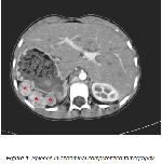
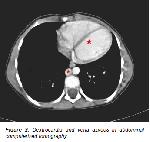
A 6-year-old boy patient was presented in to our hospital with night cough complaints. History revealed that coughing events were occasionally seen in the history of the patient during the last few months and ceased after medication. We also learned that the coughing was dry in nature. Past medical history and family history were not contributory. A physical examination indicated that the patient's general condition was good; his body weight was measured as 20 kg (25-50 percentile) and height as 120 cm (75-90 percentile). Blood pressure was 100/60 mmHg, cardiac pulse 100/minute, body temperature 36.5º C and respiratory rate was 30/minute. Oropharynx was hyperemic, cardiac pulse was at the right hemithorax and a systolic murmuring was present at 2/6 degree which was heard better at the 4-5 intercostal range of the right sternal edge. The physical examination of respiratory system and abdomen was normal. Laboratory results were as follows: urinalysis was normal. Leukocytes were 8810/mm3 and hemoglobin 13.3 gr/dL. Hepatic and renal function tests were normal. Prothrombin time and active partial thromboplastin time were within the normal range. C-reactive protein was determined to be 3 mg/dL and sedimentation was 10 mm/h. Cardiac pulse was observed at the right hemithorax and dextrocardia was detected in telecardiography. Echocardiography displayed a slight tricuspid valve failure and an isolated dextrocardia. Abdominal ultrasonography study showed that the spleen was not in its normal localization while the left lobe of the hepatic was extended to the lodge of the spleen. Multiple numbers of solid structures were observed which were considered compatible with the spleen adjuvant to the right lobe inferior to the hepatic. Results of abdominal computerized tomography indicated that the left lobe of the larger than normal hepatic passed the midline, and that the left lobe and the caudate lobe displayed a hypertrophic image. The corpus and tail sections of the pancreas could not be monitored, a heterogeneous contrasting was observed at the lodge of the spleen, the largest of which was 3 cm in size while multiple regularly bordered images compatible with the spleen were also observed (Figure 1). Vena cava inferior was not observed, it was determined that vena azygos was wider than normal and that drainage was carried out via this vein (Figure 2).
A portal vein colored doppler ultrasonography study displayed a high portal vein flow at the hylus of the hepatic while the vena cava inferior was absent and the direction of the flow was orientated towards the hepatic. The findings above supported the diagnosis of PS in this case. The patient could not be further investigated because the family requested a referral to the high center. The patient was referred to an advanced center for possible surgical intervention in the future. |
|||||
| Discussion | |||||
Polysplenia/heterotaxy syndrome is a very rare anomaly that includes gastrointestinal, vascular and cardiac malformations and may be associated with thoracic and abdominal organ malformation and dysmorphism6,7. Diagnosis is possible with cardiac complaints that are especially frequent in childhood. PS generally causes no complaints until adulthood and is most often diagnosed incidentally as a result of radiological investigations performed for other reasons5. Mortality rates are high among PS patients due to cardiac abnormalities, biliary or intestinal atresia as half of the patients may survive to be four months old while 25% of PS patients may manage to stay alive until five years of age2. Adult patients without cardiac abnormalities or with minor cardiac abnormalities may reach the adult term of their lives; however, this group of patients constitutes 10% of the entire cases2. The most frequent abnormality detected in PS is multiple numbers of spleens. The spleens are 1-6 cm in dimension, and can be located at the right or left of the abdomen while the gastric is generally adjuvant to the large curvature. The second frequent finding is vascular anomalies. The continuation of the vena cava inferior azygos or hemiazygos and the total absence of the hepatic segment are the most frequently encountered vascular anomalies and can be seen in 50-60% of the cases7,8. The problem here is the absence of the vena cava inferior suprarenal section, which drains into the superior vena cava instead of the right atrium as it shows a continuation with the expanded azygos or hemiazygos vein located at infrarenal vena cava inferior. In Polysplenia cases where a left isomerism is seen the spleen tissue is always located alongside the large curvature of the gastric5,8. In a number of eight PS cases studied by Gayer et al9, spleens were located at the adjuvant of the gastric's large curvature. In our case, spleens were observed in the left upper quadrant. Polysplenia/asplenia is a frequent abnormality that can be seen in the bilateral superior vena cava10. However, hepatic veins may directly open to the right atrium8. In our case, vena cava inferior could not be detected, but vena azygos was wider than normal and therefore drainage was possible by means of this vein. Patients with minor cardiac malformations or without any cardiac anomalies comprise 10% of all patients and these patients represent the group of patients that have achieved their adult term9. Biliary or intestinal atresia was not detected in our case; however, an isolated dextrocardia was present. However, the gastric and hepatic can be located at the right/left or midline8. In our case, no anomalies related to the hepatic or gastric were encountered. The pancreas may be located at its normal localization or at the right of the tail section. Another abnormality which can be observed in the pancreas is its shortness. Short pancreas forms due to developmental failure of the tail and corpus sections of the pancreas as a result of an undeveloped dorsal bud during the development stage of the pancreas10. In patients with a short pancreas, only the head section of the pancreas can be observed and the corpus and tail sections cannot be observed. In our case corpus and the tail sections of the pancreas were not observed. Polysplenia Syndrome is a syndrome where multiple anomalies may accompany the condition. The disease is a rare condition and may not be diagnosed alone. Knowledge of the anomalies in this syndrome can avoid erroneous diagnoses of short pancreas and interpretation of spleens as masses. This case was presented since it is a rare condition where dextrocardia, absence of the vena cava inferior and polysplenia are seen together. |
|||||
| References | |||||
1) Winer-Muram HT, Tonkin ILD. Spectrum of heterotaxic syndromes. Radiol Clin North Am 1989; 27: 1147-1170.
2) Peoples WM, Moller JH, Edward JE. Polysplenia: A review of 146 cases. Pediatr Cardiol 1983; 4: 129-137.
3) Roguin N, Hammerman H, Korman S, Riss E. Angiography of azygos continuation of inferior vena cava in situs ambiguus with left isomerism (polysplenia syndrome) Pediatr Radiol 1984; 14: 109–112.
4) Winer-Muram HT. Adult presentation of heterotaxic syndromes and related complexes. J Thorac Imaging 1995; 10: 43–57.
5) Ahmetoğlu A, Koşucu P, Sarı A, Gümele H.R. Polisipleni sendromunda radyolojik bulgular. Tanısal ve Girişimsel rad-yoloji 2002; 8: 510-512.
6) Kapa S, Gleeson F.C, Vege S.S. Dorsal pankreas agenezis and polysplenia/heterotaxy syndrome. JOP 2007; 8: 433-437.
7) Van de Pere S, Vanhoenacker F.M, Petre C, Van Doorn C, De Schepper A.M. Heterotaxy Syndrome JBR-BTR 2004; 87: 158-159.
8) Schulman M.H. Asplenia/polysplenia. Emecidine World Medical Library.
(http://emedicine.medscape.com/article/406116-overview) (Updated: Jun 17, 2009)
|
|||||
| [ Başa Dön ] [ Özet ] [ PDF ] [ Benzer Makaleler ] [ Yazara E-Posta ] [ Editöre E-Posta ] | |||||
| [ Ana Sayfa | Editörler | Danışma Kurulu | Dergi Hakkında | İçindekiler | Arşiv | Yayın Arama | Yazarlara Bilgi | E-Posta ] |

