 |
 |
| [ Ana Sayfa | Editörler | Danışma Kurulu | Dergi Hakkında | İçindekiler | Arşiv | Yayın Arama | Yazarlara Bilgi | E-Posta ] | |
| Fırat Tıp Dergisi | |||||||||||
| 2013, Cilt 18, Sayı 1, Sayfa(lar) 051-053 | |||||||||||
| [ Özet ] [ PDF ] [ Benzer Makaleler ] [ Yazara E-Posta ] [ Editöre E-Posta ] | |||||||||||
| Primary Retroperitoneal Mucinous Cystadenoma with a Sarcoma-Like Mural Nodule: Ultrasound and MRI Findings | |||||||||||
| Ahmet Kürşad POYRAZ1, Muammer AKYOL1, Ercan KOCAKOÇ2 | |||||||||||
| 1Fırat University Faculty of Medicine, Department of Radiology, Elazig, Turkey 2Bezmialem Foundation University Faculty of Medicine, Department of Radiology, Istanbul, Turkey |
|||||||||||
| Keywords: Mucinous cystadenoma, Retroperitoneum, Sarcoma, MRI, Müsinöz kistadenom, Retroperiton, Sarkom, MRG | |||||||||||
| Summary | |||||||||||
Primary retroperitoneal mucinous cystadenoma with a sarcoma-like mural nodule is a rare entity with one case reported in literature. We present a case of a 34-year-old woman who presented for evaluation of gastric pain, where a cystic abdominal mass was incidentally seen on ultrasonography. Subsequent magnetic resonance imaging (MRI) showed signal characteristics of a retroperitoneal cystic mass with hemorrhagic content and a mural nodule. After one year there was no evidence of recurrence or metastasis on follow-up ultrasonography. |
|||||||||||
| Introduction | |||||||||||
Cystic masses originating from retroperitoneum are rare. The types of retroperitoneal cystic masses are cystic lymphangioma, mucinous cystadenoma, cystic mesothelioma, epidermoid cyst, pseudomyxoma retroperitonei, mullerian cyst, tailgut cyst, bronchogenic cyst, cystic change in solid neoplasms, and cystic teratoma1. We report the imaging findings of a mucinous cystadenoma with a sarcoma like mural nodule originating from retroperitoneum. |
|||||||||||
| Case Presentation | |||||||||||
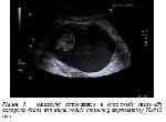
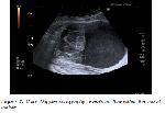
A 34-year-old woman presented for evaluation of epigastric pain and discomfort for one year. Physical examination revealed epigastric tenderness and laboratory findings were within normal limits. An initial transabdominal sonography demonstrated a large cystic mass with a mural nodule in left lower quadrant (Figure 1). Cystic mass was located anterior to the left psoas muscle with approximately 110x76 mm in largest dimension. Cyst was hypoechoic with thin wall and regular contour without septation and the mural nodule was heterogenous hypoechoic and there was no blood flow signal within the nodule in color Doppler evaluation (Figure 2).
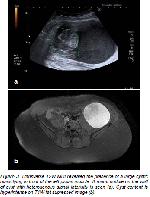
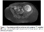
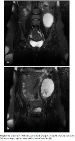
Subsequent contrast enhanced abdominal magnetic resonance imaging (MRI) was performed to evaluate nature and extent of the mass. The MRI revealed an 11x8 cm cystic retroperitoneal mass (Figure 3) with a mural nodule. The mass was located in retroperitoneal space of left lower quadrant in front of the psoas muscle. The cyst was hyperintense in both fat-suppressed T1-weighted spin echo and T2-weighted fast spin echo images. The mural nodule (Figure 4) measured approximately 5x5 cm and showed minimal enhancement in the contrast-enhanced T1-weighted fat-suppressed images. There was no enhancement in the thin wall of cyst. The surrounding organs were displaced by the mass however there was no evidence of invasion. Both ovaries and uterus were normal (Figure 5). There was no distant metastasis seen on abdominal MRI. Serum CA-125 and CA-15-5 levels were within normal limits however CA-19.9 level was elevated (115 U mL).
The patient underwent laparotomy and a retroperitoneal capsulated cystic mass was confirmed. The tumor weighed 470 g and measured 9×10×14 cm. The histological examination described hemorrhagic content in the cystic space and a mural nodule measuring 5 cm in diameter. Diagnosis was mucinous cystadenoma with sarcoma like mural nodule. The mural nodule was positive for vimentine and CD68 and negative for pancytokeratin, CD10 and ER. Twelve months following the initial surgical resection there was no evidence of recurrence or metastasis on follow-up ultrasonography. |
|||||||||||
| Discussion | |||||||||||
Primary retroperitoneal mucinous cystadenoma with mural nodule is an extremely rare tumor. To our knowledge this is the first report demonstrating the ultrasonography and MRI findings of a primary retroperitoneal cystadenoma with a sarcoma-like mural nodule. Imaging findings of primary retroperitoneal mucinous cystadenomas are similar to ovarian mucinous cystadenomas. These tumors occur in women with normal ovaries. Delay in the diagnosis of retroperitoneal tumors is common due to nonspecific symptoms. Transabdominal ultrasound, computed tomography (CT) and MRI can help diagnosis and reveal the nature of the retroperitoneal cysts however histologic examination is required for the definite diagnosis. Primary mucinous tumors of the retroperitoneum can be classified into three clinicopathologic types: mucinous cystadenoma, mucinous cystic tumor of borderline malignancy, and mucinous cystadenocarcinoma2. Mural nodules of mucinous ovarian cysts include: anaplastic carcinomas, sarcomas, carcinosarcoma, sacoma-like nodules, mixed nodules and leiomyomas2-4. Ovarian mucinous cystadenomas with sarcoma-like mural nodules has been described in a few reports however sarcoma-like nodules in primary retroperitoneal mucinous cystsadenoma has been reported in only one case previously. Ultrasound images of the cyst can vary because of the spectrum of contents. Cyst content was hypoechoic due to hemorrhagic content in our case. In large cysts it can be difficult to determine the origin and extension with ultrasonography and a subsequent examination might be needed. CT can determine presence of calcifications and show the relationship of the mass with surrounding tissues. Presence of invasion or displacement of organs can be determined with CT however radiation exposure to the patient and limited soft tissue contrast are the limitations of CT. MRI is more sensitive to determine the mucinous content of cyst and enables more detailed information about mural nodule. Presence of invasion and relation with surrounding tissues can be reliably determined with MRI. The tumor was first determined by ultrasonography in our case. Our initial diagnosis was cystic malignant fibrous histiocytoma. Presence of enhancement, nature of cyst content and relation with the surrounding organs were evaluated by subsequent MRI. Preoperative diagnosis of primary retroperitoneal mucinous cystadenoma is difficult because there are no reliable markers or methods for mucinous cystadenomas5 and it should be considered as a rare differential diagnosis of an abdominal cystic mass. |
|||||||||||
| References | |||||||||||
1) Yang DM, Jung DH, Kim H, et al. Retroperitoneal cystic masses: CT, clinical, and pathologic findings and literature review. Radiographics 2004; 24: 1353-65.
2) Bakker RF, Stoot JH, Blok P, Merkus JW. Primary retroperitoneal mucinous cystadenoma with sarcoma like mural nodule: a case report and review of the literature. Virchows Arch 2007; 451: 853-7.
3) Prat J. Pathology of the ovary. Philadelphia, PA: Saunders, 2004: 123-8.
|
|||||||||||
| [ Başa Dön ] [ Özet ] [ PDF ] [ Benzer Makaleler ] [ Yazara E-Posta ] [ Editöre E-Posta ] | |||||||||||
| [ Ana Sayfa | Editörler | Danışma Kurulu | Dergi Hakkında | İçindekiler | Arşiv | Yayın Arama | Yazarlara Bilgi | E-Posta ] |
